

Welcome!
Welcome to the new CancerGRACE.org! Explore our fresh look and improved features—take a quick tour to see what’s new.
Dr. Jed Gorden, Swedish Cancer Institute, describes the differences between bronchoscopy and endobronchial ultrasound, highlighting the advantages of EBUS in diagnosis and staging.
Transcript
Today I’m going to talk to you about bronchoscopy with endobronchial ultrasound. It’s an interesting technology — bronchoscopy in its form that we know it now, flexible bronchoscopy, has been around since about the late 1960s. This allowed us to move bronchoscopy from an operating room into outpatient settings and allow us to navigate through the airways. For those of you that have heard of colonoscopies or upper endoscopies, these are all cameras that allow us to snake through the airway or other orifices and get a much clearer picture of what’s going on inside. The challenge though with traditional bronchoscopy is it allows you to only see what’s directly in front of you — what is in the airway, and the overwhelming majority of the time the airways are normal.
A tremendous advantage to us in the field of lung cancer, lung cancer diagnosis, and lung cancer staging has been bronchoscopy coupled with ultrasound, creating what’s called endobronchial ultrasound or EBUS. This is a very small ultrasound probe coupled at the end of a bronchoscope. It enters into the airway in the exact same fashion that bronchoscopy does for traditional procedures, but what it allows us to do is look through the airway wall. This is critical because looking through the airway wall now allows us to identify lymph nodes, abnormalities that are around the trachea, the bronchi which are the divisions of the airway that go to the left lung and the right lung, and this is critically important because we know that with staging where we’re not only diagnosing those that have cancer, but where the cancer is and whether it has spread to the lymph nodes is crucial to an understanding and developing a treatment plan.
Some we now have a tool in our armamentarium to, in a very minimally invasive way, go into the airways, see what’s in the airways, and see through the airways into the lymph nodes that live in and around those airways. Once we’ve identified these very specific structures, we can sample them with small needles allowing us to puncture through the airway wall directly into a lymph node, collect a sample, have a pathologist look at it under a microscope, and tell us whether that lymph node is involved in cancer or that lymph node is not involved with cancer. Critical are the decisions that will be made for creating a treatment algorithm.
The advantage of this is that it’s minimally invasive; it’s done in the outpatient setting. It allows us to sample most of the lymph nodes that are present and are critical to decision making around lung cancer and lung cancer staging. Complications of it are very rare — sometimes after bronchoscopy and bronchoscopy with ultrasound, people can experience a fever, or maybe a sore throat, but larger complications like bleeding and infection are very rare.
The most important thing though to understand is that this is a partnership with your physician and that they explain to you what procedure you’re going to have, and how this procedure is going to benefit you, whether it’s bronchoscopy or bronchoscopy with ultrasound.
The final thing that I’m going to talk about with bronchoscopy and bronchoscopy with ultrasound is how you’ll be during the procedure. Most patients ask, “am I going to be awake; am I going to know what’s going on?” There are two ways to do bronchoscopy and bronchoscopy with ultrasound. One is what’s called conscious sedation — this is sort of a twilight phase where people are sleeping, they’re breathing on their own, responsible for their own vital signs, but a bronchoscopist is allowed to do procedures without it causing too much disturbance to the patient. This is good for procedures that last about 25 to 30 minutes and allows people to sample in the airways in a very safe fashion. Another way that bronchoscopy with ultrasound is performed is with anesthesia — this is where an anesthesiologist takes over the safety of the patient and the control of their airway. A breathing tube or a small cap over the back on the airway is placed allowing air to pass in and out and control the breathing and ensure safety during the procedure.
So when you talk with your physician about this, it’s important to understand how you will feel during the procedure, what is going to be going on in terms of your safety, sedation, and your comfort. It’s also important to know that there’s data for this; the data for this suggests that the procedures are equal. Bronchoscopy and bronchoscopy with ultrasound can be done safely in the setting of conscious sedation and in the setting of general anesthesia, and you should feel confident that you can have a safe and effective procedure.
So in summary the most important thing is that you partner with your physician in order to get the most information possible from any procedure. In this case the procedure will be bronchoscopy. Bronchoscopy is an inspection of the airway. We couple that with ultrasound which is not only inspection of the airway, but visualization through the airway wall, identifying the lymph nodes and structures. Biopsying those gets tissue which is staging and telling us how much cancer there is, and this can be done safely and effectively with you sleeping in either a conscious or twilight phase, or with general anesthesia.
Please feel free to offer comments and raise questions in our
discussion forums.


Dr. Singhi's reprise on appropriate treatment, "Right patient, right time, right team".
While Dr. Ryckman described radiation oncology as "the perfect blend of nerd skills and empathy".
I hope any...
My understanding of ADCs is very basic. I plan to study Dr. Rous’ discussion to broaden that understanding.
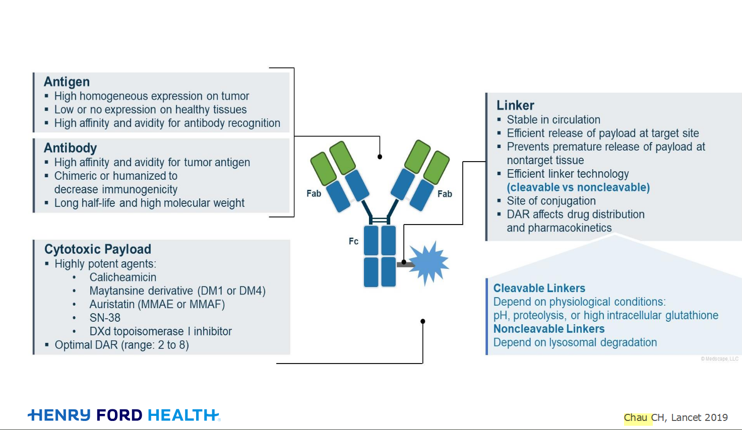
An antibody–drug conjugate (ADC) works a bit like a Trojan horse. It has three main components:
Bispecifics, or bispecific antibodies, are advanced immunotherapy drugs engineered to have two binding sites, allowing them to latch onto two different targets simultaneously, like a cancer cell and a T-cell, effectively...
The prefix “oligo–” means few. Oligometastatic (at diagnosis) Oligoprogression (during treatment)
There will be a discussion, “Studies in Oligometastatic NSCLC: Current Data and Definitions,” which will focus on what we...
Radiation therapy is primarily a localized treatment, meaning it precisely targets a specific tumor or area of the body, unlike systemic treatments (like chemotherapy) that affect the whole body.
The...

Welcome to the new CancerGRACE.org! Explore our fresh look and improved features—take a quick tour to see what’s new.
A Brief Tornado. I love the analogy Dr. Antonoff gave us to describe her presentation. I felt it earlier too and am looking forward to going back for deeper dive.