Welcome!
Welcome to the new CancerGRACE.org! Explore our fresh look and improved features—take a quick tour to see what’s new.
General Introduction to Small Cell Lung Cancer Lung cancer consists of two major types: small cell lung cancer (SCLC) and non-small cell lung cancer (NSCLC). Approximately 85% percent of all lung cancer patients have NSCLC, and the remaining 15% have SCLC.  (click on image to enlarge) In 2010, the American Cancer Society has estimated that approximately 222,000 new cases of lung cancer will be diagnosed, of which 35,000 will have SCLC. Even though both subtypes are lung cancers, they are considered as separate diseases in most ways, and the management of these two cancers is different. It is important to recognize that the treatments applicable for NSCLC, including many newer agents that have been approved and are the subject of increasing research and media attention, are not clearly relevant for patients with SCLC.
Symptoms and Work up Nearly all the patients with SCLC have symptoms at the time of diagnosis. The common symptoms that many patients have at that time are chest pain, cough, and shortness of breath. Some patients may note fatigue and weight loss at the time of initial presentation.  Patients may also have symptoms related to the sites to which the cancer has spread. For example, patients may develop headache or blurring of vision due to spread to the brain, abdominal pain from spread to the liver, or focal bone pain from spread to the bones. However, we often find that patients have tumor spread to areas of the body for which they do not have any specific symptoms. Importantly, patients generally have symptoms for only a short period of time before diagnosis. This is related to the relatively rapid growth of SCLC. Symptoms related to Paraneoplastic Syndromes- Paraneoplastic syndromes are changes in different body parts that can occur in cancer patients, not related to direct spread of the cancer. Almost 50% of SCLC patients could have one or more paraneoplastic syndromes, with symptoms that often precede the diagnosis of SCLC. Some of the common paraneoplastic syndromes are listed in the table below.
  Many symptoms from the paraneoplastic syndromes improve with treatment, but this does not always occur. Worsening of the symptoms from paraneoplastic syndromes typically indicates progression of cancer. Staging of Patients with SCLC The purpose of staging is to find out 1. What is the extent of SCLC? In other words, where the cancer has spread to, since the extent of the cancer determines the treatment 2. What is the general condition of the patient? The following tests are done in patients who are diagnosed with SCLC or suspected to have SCLC:
Note that PET scans are not an approved test for SCLC, even though it is approved for NSCLC (due to it being better studied in that setting). However, if a PET scan is done, then a bone scan is not needed, but a brain scan is still required, since the brain is not well examined on a routine PET scan. These studies are routinely performed because the areas in the body to which SCLC most typically spreads include the lymph nodes within the chest (hilar: lymph nodes within the lungs, and mediastinal lymph nodes: lymph nodes in the middle of the chest), the brain, other portions of the lung or the opposite lung, pleura (lining of the lungs), adrenal glands, liver, and bones (see figure below).  It is important to recognize, however, that SCLC can spread to any other part of the body, such as the kidneys. It is also important to recognize that many patients have SCLC that has not spread widely throughout the body. Unfortunately, even if the scans of the different body parts reveal no evidence of cancer, we cannot rule out that there is cancer involvement of these areas; rather, we can only safely conclude that if the cancer has spread to the other parts in the body, it has not grown sufficiently big enough for it to be seen on the scans. Small Cell Lung Cancer Stages Generally, every cancer patient's cancer is assigned a stage. The stage of the cancer represents the extent of the spread of the cancer as can be detected with physical exam, blood tests, and scans. In lung cancer, both NSCLC and SCLC, there are four stages. In stage I and II, the lung cancer is detected only in the lung or in the lung and in the hilar lymph nodes. In stage III, the lung cancer is detected not only in the lung, but also there is evidence that the cancer has spread to the mediastinal lymph nodes. In stage IV, spread of lung cancer can be detected in other parts of the body. Spread to the opposite lung also represents stage IV lung cancer. The most important features of SCLC are its proclivity to spread to other parts of the body early in the course of the cancer and its ability to grow relatively rapidly. Due to its ability to spread to other parts of the body early, we recommend chemotherapy even for earlier stage SCLC based on recognized risk that the cancer has at least microscopic spread (micrometastases), even if it is not detected in the other parts of the body on any of the scans. Traditionally small cell lung cancer has been grouped into one of 2 stages: limited or extensive stage. Limited Stage: SCLC is considered to be limited if the cancer is detected only in the lung itself, or the lung and in the hilar or mediastinal lymph nodes. In terms of the stages mentioned earlier, limited stages consists of stage I, II and III.
  About 25-30% of all patients with SCLC have limited disease at the time of diagnosis. Extensive Stage; SCLC is considered extensive disease (ED) if the spread of the cancer can be detected in other parts of the body. In terms of the stages mentioned earlier, extensive stage consists of stage IV. The areas of the body that SCLC commonly spreads to are the brain, bones, liver, adrenal glands, or other parts of the lungs. Another important area of spread is the pleura. The pleural lining is a thin wrap (like a saran wrap) that surrounds each of the two lungs and is normally too thin to be seen on scans. The wrap doubles on itself and forms a pleural cavity between the two folds of the wrap. The inner fold of the wrap is called the visceral pleura, while the outer fold is called the parietal pleura, as shown in the figure below.
  Normally, this cavity has a tiny amount of fluid. Please note that the image shown above shows a pleural cavity that is far larger than normal for easier illustration. Spread of the cancer to the pleura usually leads to formation of excess fluid in the pleural cavity. Accumulation of fluid in the pleural cavity, as shown in the illustration below, compresses the lung and leads to shortness of breath and sometimes a cough.
  Even if the spread in a patient is only observed in the pleura, or cancer cells are seen within the pleural fluid, the patient is generall considered to have ED-SCLC. Spread to the pleura is not a result of direct extension of the cancer to the pleura, but spread through the blood stream. Thus if it has spread to the pleura, it demonstrates the presence of micrometastatic SCLC cells in the blood stream. This also means that SCLC cells have also reached some other parts of the body, though this is presumably not seen only because evidence of cancer in these other parts of the body has not grown big enough to be seen on the scans. Data show that survival of patients is better for patients who have disease limited only to the pleura or pleural effusion compared with more widespread metastatic disease. About 70-75% of all patients with SCLC have extensive disease at the time of diagnosis. Treatment of SCLC will be covered in a separate summary chapter. The GRACE Lung Cancer Reference Library is made possible by an unrestricted educational grant from Pfizer Oncology.
Please feel free to offer comments and raise questions in our
discussion forums.
Hi app.92, Welcome to Grace. I'm sorry this is late getting to you. And more sorry your mum is going through this. It's possible this isn't a pancoast tumor even though...
A Brief Tornado. I love the analogy Dr. Antonoff gave us to describe her presentation. I felt it earlier too and am looking forward to going back for deeper dive.
Dr. Singhi's reprise on appropriate treatment, "Right patient, right time, right team".
While Dr. Ryckman described radiation oncology as "the perfect blend of nerd skills and empathy".
I hope any...
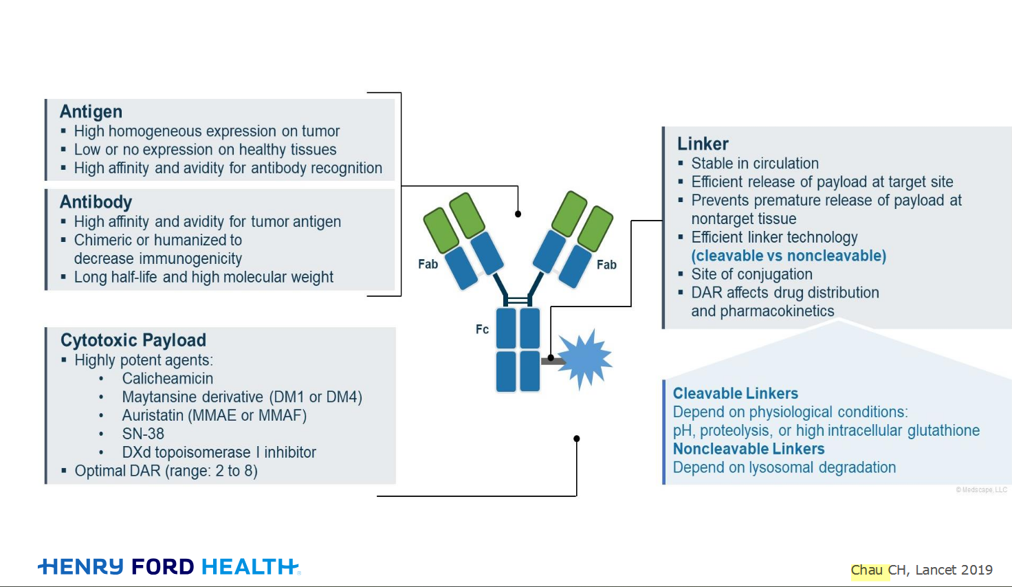
My understanding of ADCs is very basic. I plan to study Dr. Rous’ discussion to broaden that understanding.

Here's the webinar on YouTube. It begins with the agenda. Note the link is a playlist, which will be populated with shorts from the webinar on specific topics
An antibody–drug conjugate (ADC) works a bit like a Trojan horse. It has three main components:

Welcome to the new CancerGRACE.org! Explore our fresh look and improved features—take a quick tour to see what’s new.