Welcome!
Welcome to the new CancerGRACE.org! Explore our fresh look and improved features—take a quick tour to see what’s new.
One subtype of lung cancer that we haven’t specifically talked about is called a Pancoast tumor, named for the doctor who first described them. A Pancoast tumor is a NSCLC that is located in a groove called the superior sulcus (Pancoast tumors are also sometimes referred to as superior sulcus tumors), at the top (or apex) of each of the lungs. Here's the appearance of one on a chest x-ray:
(Click to enlarge)
Typically squamous cancers, they are located high up in the chest and often very close to or actually growing into the chest wall or vertebrae, making it very hard or potentially impossible to remove them surgically. In fact, they were initially described back in the 1950s as unresponsive to radiation, unresectable, and therefore 100% fatal within a couple of years. Fortunately, we have made steady progress and now definitely feel that these tumors are potentially curable, with the evidence now to prove that.
The first baby steps described individual cases of patients who lived for several years after radiation (chemotherapy and the field of medical oncology really were just in their infancy at that time, and this far preceded the time of any effective chemo for lung cancer). A key publication (Haas, JAMA, 1954, too old for an online abstract) was titled, "Radiation Management of Otherwise Hopeless Neoplasm" (yikes!) and described a patient who lived for nearly three years after radiation. A few other encouraging cases trickled in of long-term survivors. In 1956, a report was published (Shaw and colleagues, Annals of Surgery) of a patient received three weeks of radiation and then, three weeks later, went to another institution to see a surgeon who decided to try to resect the cancer. The surgeon noted that the surgery went smoothly, and that the cancer was dead on the periphery but viable in the center. The patient remained alive for more than 5 years afterward. While it was only one patient, it proved what was possible.
And then, based on that experience of one man with a Pancoast tumor happening to go to see a surgeon three weeks after completing radiation and having encouraging early results, the approach of waiting three weeks after radiation and then attempting surgery for similar cases became the thing to do. These were still unusual to rare cases, but a few small studies emerged describing several long-term survivors, including one study in 1975 (abstract here) from 61 resected patients over 19 years at Baylor who were all treated with radiation followed by surgery, with a 5-year survival of 35%. Not as high we we’d like, but much better than the assessment that this was a "hopeless neoplasm" 20 years earlier.
The Modern Era
Although Pancoast tumors are still challenging to treat, we’ve got several new tools, including much better imaging techniques, and chemotherapy. First, the value of newer radiologic tests like CT and PET scans, can provide much better detail of where the cancer is and what it is or isn’t invading:
Precise imaging is particularly helpful for radiation oncologist who need to determine exactly what to radiate and what normal tissue to spare, and for surgeons to try to anticipate what they will see and whether the tumor is invading into the vertebrae or other areas that may be very difficult or impossible to remove surgically. And as with PET scans for other lung cancers, they can also detect whether the cancer has spread to other areas that might make surgery inappropriate, such as if metastatic spread to the liver, adrenal glands, etc. is observed. While Pancoast tumors are typically squamous and tend to grow more locally than spread distantly, we certainly see metastatic spread in some patients and wouldn’t want to perform a surgery if it wouldn’t provide a realistic hope of significant benefit. At my institution, for these patients we routinely obtain CT and PET imaging as part of our standard staging workup, as well as a brain MRI, looking for distant spread before undertaking surgery.
I’m not a surgeon, so I won’t go into much detail on the evolution of surgical approaches, but they’ve made advances over the past 15-20 years. The older approach was with a large incision from the back:
But 10-15 years ago, two thoracic surgery groups from France pioneered new approaches from the neck, with the incision either cutting through (abstract here) or below the clavicle (extract here):
These new techniques can allow for greater visibility and access to many Pancoast tumors, particularly those toward the anterior, or front, part of the chest:
Next we’ll turn to the trimodality approach that includes induction chemo and radiation all followed by surgery, which is now what many would considered our current best treatment approach for patients who can pursue it.
Please feel free to offer comments and raise questions in our
discussion forums.
Dr. Singhi's reprise on appropriate treatment, "Right patient, right time, right team".
While Dr. Ryckman described radiation oncology as "the perfect blend of nerd skills and empathy".
I hope any...
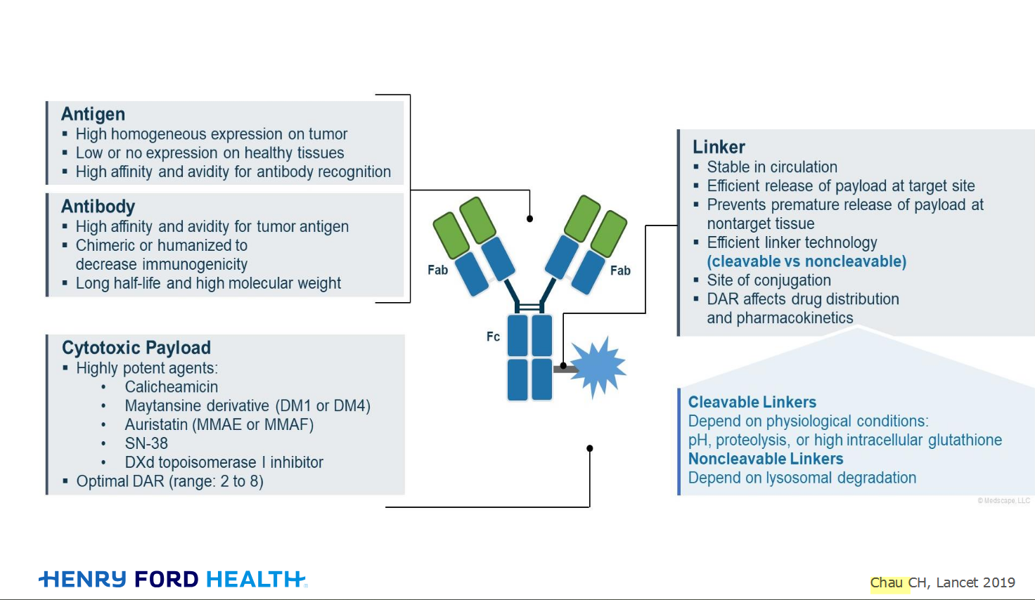
My understanding of ADCs is very basic. I plan to study Dr. Rous’ discussion to broaden that understanding.

An antibody–drug conjugate (ADC) works a bit like a Trojan horse. It has three main components:
Bispecifics, or bispecific antibodies, are advanced immunotherapy drugs engineered to have two binding sites, allowing them to latch onto two different targets simultaneously, like a cancer cell and a T-cell, effectively...
The prefix “oligo–” means few. Oligometastatic (at diagnosis) Oligoprogression (during treatment)
There will be a discussion, “Studies in Oligometastatic NSCLC: Current Data and Definitions,” which will focus on what we...
Radiation therapy is primarily a localized treatment, meaning it precisely targets a specific tumor or area of the body, unlike systemic treatments (like chemotherapy) that affect the whole body.
The...

Welcome to the new CancerGRACE.org! Explore our fresh look and improved features—take a quick tour to see what’s new.
A Brief Tornado. I love the analogy Dr. Antonoff gave us to describe her presentation. I felt it earlier too and am looking forward to going back for deeper dive.