Welcome!
Welcome to the new CancerGRACE.org! Explore our fresh look and improved features—take a quick tour to see what’s new.
When oncologists and surgeons talk about staging, we often distinguish between clinical and pathologic staging. Many in the health care field don’t understand or know the difference. Even more, why do we “stage” a cancer (NOT the patient!) at all? These are important questions, because they tell those of us involved in the treatment and care of such patients what is the extent of the disease, what the prognosis might be, and what the treatment plan should entail. That way, the caregivers are all “on the same page".
It is quite important to know that staging is done, by tradition, ONLY at the discovery of the disease and a tissue diagnosis (by biopsy) obtained, and before the start of treatment. When and if there is a recurrence of the cancer, we do not give the cancer a new stage. It is then just that, a recurrence. Thus, the term “re-staging” is really a misnomer; the process would be better termed a “re-evaluation”. Another common misconception in staging and diagnosis among patients is related to the spread of the cancer from one organ to another: if a lung cancer has spread to the bone or liver, it's still one process. It isn't called a bone or liver cancer, but rather, it is lung cancer metastatic to those places.
Focusing on non-small cell lung cancer (NSCLC), for instance, clinical staging refers to what we know after all our tests have been performed, such as physical examination, chest x-ray, CT scan (usually of the chest and upper abdomen, the latter to look for involvement of the liver, adrenal glands, and lymph nodes), total body PET scan, and, ideally, MRI of the brain (PET is negative 40% of the time when there is spread to the brain; also, CT scans of the brain are not as sensitive and accurate as MRIs). Bone scans rarely need to be performed anymore, having been replaced by PET.
At this point, we turn to the status of the mediastinum, the mid-chest area between the two lungs, where some important lymph nodes that drain from lungs are located. The importance here is to determine, before any surgery, that these nodes do not contain cancer; if involved, they are designated as N2 nodes if they're on the same side as the cancer, and N3 nodes if they're on the opposite side from the cancer (N1 nodes are within the same lung as the cancer). Most experts do not favor immediate surgery in most cases if these nodes are “positive” for cancer involvement. Instead, surgery is rarely employed at all for patients with N3 nodes, and it's hotly debated if and when it should be done for patients with N2 nodes.
When and how is the mediastinum evaluated? If the CT/PET do not suggest involvement (“negative” study), sometimes a patient will go straight to surgery, but it's also not rare for there to be cancer involved when the imaging looks pretty unremarkable (a "false negative" study). If the surgeon wants a higher level of certainty, the next step would be what is called a “mediastinoscopy". This is a procedure in which the surgeon looks with a scope into the mediastinum and obtains biopsies. In some situations the surgeon may elect, instead, to open the chest to the side of the breast bone (sternum), depending on the location of the cancer and the suspected lymph nodes. This is sometimes referred to as a “Chamberlain procedure”. In addition, a relatively recent development that represents a less aggressive procedure is an endoscopic ultrasound down through the esophagus and/or large airway (bronchus). This effort provides a potential for a less invasive approach to obtaining tissue from nodes or sometimes a central tumor mass.
At this point, clinical staging is complete. Next comes the decision as to whether to operate for potential cure, and what type of surgery should be performed. At the time of surgery, the decision will be made as to whether a wedge resection, segmental resection, lobectomy, or pneumonectomy (the entire lung on that side) will be performed. The highest priority is that all of the tumor must be removed, with some uninvolved lung tissue surrounding it. Importantly, the mediastinal lymph nodes on that side (N2) should also be removed. The current preferred standard is for at least 12 nodes to be removed and labeled as to location, except, perhaps for some very small tumors.
When this surgery is complete, we will have the pathologic staging, which is, of course, more accurate and definitive than clinical staging (note that pathologic staging is only defined after surgery, not after sampling areas with limited biopsies). Sometimes pathologic stage turns out, in fact, be lower than clinical stage, such as if a patient has enlarged lymph nodes from infection along with a cancer, but those nodes aren't involved with the cancer itself.
After recovery from surgery the decision will be made as to need for chemotherapy and/or radiation therapy. Staging of NSCLC is frequently changing, as we learn more and try to “fine tune” the process. Importantly, though, the current staging process includes tumor size and extent of spread but doesn't yet include refinements by molecular characteristics. Most of us in the field consider this to be the next major step in refining prognosis and treatment plans for our patients with cancer.
Please feel free to offer comments and raise questions in our
discussion forums.
Hi app.92, Welcome to Grace. I'm sorry this is late getting to you. And more sorry your mum is going through this. It's possible this isn't a pancoast tumor even though...
A Brief Tornado. I love the analogy Dr. Antonoff gave us to describe her presentation. I felt it earlier too and am looking forward to going back for deeper dive.
Dr. Singhi's reprise on appropriate treatment, "Right patient, right time, right team".
While Dr. Ryckman described radiation oncology as "the perfect blend of nerd skills and empathy".
I hope any...
My understanding of ADCs is very basic. I plan to study Dr. Rous’ discussion to broaden that understanding.

Here's the webinar on YouTube. It begins with the agenda. Note the link is a playlist, which will be populated with shorts from the webinar on specific topics
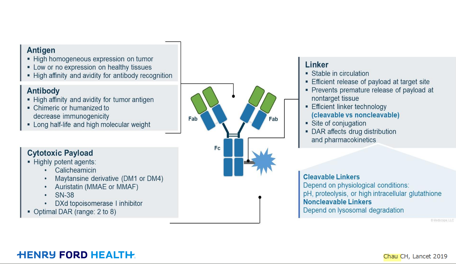
An antibody–drug conjugate (ADC) works a bit like a Trojan horse. It has three main components:

Welcome to the new CancerGRACE.org! Explore our fresh look and improved features—take a quick tour to see what’s new.