Welcome!
Welcome to the new CancerGRACE.org! Explore our fresh look and improved features—take a quick tour to see what’s new.
A publication by Drs. Oxnard and colleagues from Memorial Sloan-Kettering Cancer Center just came out in the Journal of Clinical Oncology that should remind all of us of the pitfalls of taking very small changes in measurements too literally. As an exercise to test the variability in CT scanning, they had 30 eligible patients with stage III or IV lung cancer and a minimum tumor size of 1 cm undergo a CT scan, wait a few minutes, and then have a repeat CT scan within less than 15 minutes on the same scanner. Any of 3 expert radiologists read the scans and did tumor measurements, blinded to the timing of the scans. What they found was that 57% of the patients had a measurable difference in tumor size of a mm or more, and a third had a detected difference of 2 mm. The range of difference was as dramatic as a 23% shrinkage or 31% enlargement between the two scans, while 16% had a change of more than 10%, and 3% had a change significant enough within less than 15 minutes to be meet the criteria for disease progression by the formal criteria we use to assess response. Moreover, you could put these differences on a "waterfall plot" (with relative progression plotted as bars going up on the left side and relative shrinkage as bars going down on the right side of the curve) and it would produce a range of activity that would produce a fair amount of excitement that we'd found a new treatment that helps many of our patients:  (click on image to enlarge)
Now, let's put aside the fanciful idea that the cancer has actually grown in less than 15 minutes, or that the effect of the radiation from a scan a few minutes ago led to tumor shrinkage measured on a repeat scan after a brief coffee break (or that the coffee shrank the tumor). Instead, what this should underscore is that tumor measurements aren't perfect. First, you can have slightly different appearance from one scan to the next because the slices of the scans can capture the nodule/mass at levels of slightly different thickness (imagine cutting 1-2 cm thick slices of a tennis ball: you might get different sizes depending on whether you catch the dead center of the ball or just on either side of it). More significantly, and likely far more commonly, you can get small differences based on the measurements of one radiologist vs. another or even the same radiologist reviewing a scan at two different times. There is a human element in the interpretation, which means that there is room for variability. This should also put the question of significance of differences of millimeters in measuring tumor progression into better perspective! I have long discussed the distinction between perceptible and clinically significant progression. Doctors and patients alike often over-react to incredibly minimal changes in CT scans, sometimes having people discontinue an otherwise very effective and well tolerated therapy because a new scan shows a millimeter or two of interval progression. We should bear in mind that you can see that level of variability just in redoing the scan a few minutes later or having it re-read by someone else, or perhaps the same radiologist after they have a cup of coffee. This is the same reason that many of the experts here suggest exercising caution in making broad interpretations of the significance of a slight bump in a serum tumor marker like CEA or an increase in the metabolic activity of a spot on a PET scan; these latter examples are less validated as ways to follow a cancer than a CT scan, and yet even here we see that our best and most objective criteria can fail us far more often than we'd suspect. So while this is humbling, the theme is to put observed changes in perspective of the bigger picture. A tiny change may just be noise in the system rather than a reason to reflexively jump to the next treatment.
Please feel free to offer comments and raise questions in our
discussion forums.
Dr. Singhi's reprise on appropriate treatment, "Right patient, right time, right team".
While Dr. Ryckman described radiation oncology as "the perfect blend of nerd skills and empathy".
I hope any...
My understanding of ADCs is very basic. I plan to study Dr. Rous’ discussion to broaden that understanding.

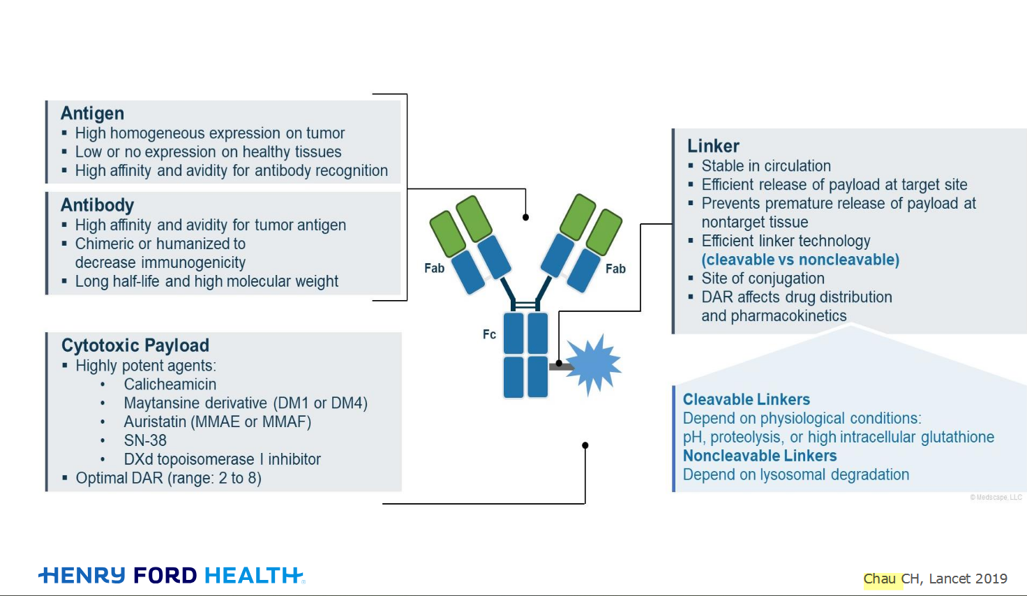
An antibody–drug conjugate (ADC) works a bit like a Trojan horse. It has three main components:
Bispecifics, or bispecific antibodies, are advanced immunotherapy drugs engineered to have two binding sites, allowing them to latch onto two different targets simultaneously, like a cancer cell and a T-cell, effectively...
The prefix “oligo–” means few. Oligometastatic (at diagnosis) Oligoprogression (during treatment)
There will be a discussion, “Studies in Oligometastatic NSCLC: Current Data and Definitions,” which will focus on what we...
Radiation therapy is primarily a localized treatment, meaning it precisely targets a specific tumor or area of the body, unlike systemic treatments (like chemotherapy) that affect the whole body.
The...

Welcome to the new CancerGRACE.org! Explore our fresh look and improved features—take a quick tour to see what’s new.
A Brief Tornado. I love the analogy Dr. Antonoff gave us to describe her presentation. I felt it earlier too and am looking forward to going back for deeper dive.