Welcome!
Welcome to the new CancerGRACE.org! Explore our fresh look and improved features—take a quick tour to see what’s new.
I've described mediastinoscopy as a "gold standard" preoperative procedure in patients who are candidates for surgery. Although it's controversial whether patients with a very low likelihood or a very high likelihood of cancer in the mediastinal nodes (mid-chest, between the lungs) need to have this confirmed by obtaining tissue to review under a microscope, we strongly prefer to get tissue for patients in whom this is a reasonably open question. For patients who are good surgery candidates and in whom there is no evidence of cancer in the lymph nodes of the mediastinum, surgery is the preferred next step. For patients who are good surgery candidates but have lymph node involvement confirmed on the same side as the main cancer (N2 nodal involvement), we most commonly recommend chemo and sometimes radiation before pursuing surgery, or recommend chemo and radiation without surgery. And for patients who have cancer in the mediastinal lymph nodes on the side opposite the main tumor (N3 nodal disease), we generally recommend chemo and radiation rather than a surgical approach. Why the difference in management? Because as nodal stage increases, the risk of cancer recurrence outside of the chest becomes greater, meaning that we need to think about distant disease (and chemo to cover it) more and more, because even excellent local treatments like surgery or radiation, or both, just can't address micrometastatic disease outside of the treatment area.
While the gold standard has been mediastinoscopy, it's invasive and requires general anesthesia. Serious complications are rare, but it's not a trivial procedure either. And it's very difficult to do a repeat mediastinoscopy to assess response to induction (pre-operative) chemo or chemo/radiation if a prior mediastinoscopy was done for initial staging. So having another option for getting tissue from mediastinal lymph nodes, especially if it is minimally invasive, would be very welcome.
One of these options that is gaining traction uses endoscopy, a tiny camera on the end of a tube, which can be used to look down the esophagus at the stomach ("upper endoscopy" or EGD, for esophagogastroduodenography), or the bronchial tree of the lungs (bronchoscopy). These procedures usually just require sedation, don't require any incisions, and don't leave visible scars. An endoscope can have an ultrasound probe attached to it so that a trained physician can get an idea of the shape and size of things behind visible barriers, allowing them to find enlarged lymph nodes that may be behind the walls of the esophagus or bronchi. The endoscope also includes a needle that can be injected into a suspicious node or tumor within the bronchus.
Putting it all together, you have endoscopic ultrasound (EUS) for sampling tissues after looking down the esophagus, and endobronchial ultrasound (EBUS) for looking through the bronchial tree and collecting tissue from tumors and lymph nodes around the mid-chest.
Although these techniques have really been available through a very limited number of well-trained specialists at a few centers, the newer generation of pulmonologists are often trained in EBUS, so we'll probably see that available more and more. We're also seeing a growing number of publications highlighting the potential value of these techniques. For instance, the widely read Journal of the American Medical Association, or JAMA recently published a report from Dr. Wallace, a pulmonologist from the Mayo Clinic in Jacksonville, FL (abstract here). In this report, 150 patients with suspected lung cancer were enrolled over two years and underwent a combination of EUS, EBUS, and transbronchial ultrasound (conventional bronchoscopy) were done by specialists "blinded" to the results of other tests or the results of imaging studies (but not blinded during the procedure, fortunately). Of the 138 patients ultimately included and eligible, 37% didn't actually have cancer. A wide range of lung cancer subtypes, as well as an occasional lymphoma, metastatic breast cancer, or other cancer were also seen. But the important part was that EBUS with fine needle aspiration (EBUS-FNA) detected 69% of cancerous lymph nodes, while the old standard of transbronchial needly aspiration (TBNA) only detected 36%, a highly significant difference. And combining EUS and EBUS picked up more lymph nodes and was particularly sensitive, better than either approach alone.
What do these results mean? These approaches don't completely replace mediastinoscopy, but the authors pointed out that if mediastinoscopy was only done in the cases in which these endoscopic approaches were negative, it would avoid mediastinoscopy 28% of the time. And even if you didn't do any mediastinoscopies, 97% of the cases that were called negative after EUS and EBUS would have been found to be truly negative.
In truth, EUS and EBUS are better at evaluating the lymph nodes in the lower part and the back of the mediastinum, while mediastinoscopy is best at finding involved lymph nodes in the upper and front parts of the mediastinum. So depending on the CT imaging, it may be possible to target the best approach for a particular case, and sometimes these endoscopic approaches will be the best or only way to reach difficult mediastinal nodes. Another setting in which EUS and EBUS may be especially helpful is before planned induction therapy for locally advanced NSCLC. Confirming suspected mediastinal node involvement with an initial EBUS allows us to "save" the mediastinoscopy so that it can be done without scar tissue from a prior one, allowing the best assessment of whether there are still cancerous lymph nodes there after pre-operative treatment. We know that this is a very important factor, enough that many experts recommend against proceeding with surgery if the post-treatment mediastinoscopy shows involved lymph nodes.
While EBUS and EUS are still only available in a limited subset of centers, these are certainly valuable approaches, and we'll likely see them integrated more and more to standard work-up algorithms as more and more specialists become trained to do them.
Please feel free to offer comments and raise questions in our
discussion forums.
Hi app.92, Welcome to Grace. I'm sorry this is late getting to you. And more sorry your mum is going through this. It's possible this isn't a pancoast tumor even though...
A Brief Tornado. I love the analogy Dr. Antonoff gave us to describe her presentation. I felt it earlier too and am looking forward to going back for deeper dive.
Dr. Singhi's reprise on appropriate treatment, "Right patient, right time, right team".
While Dr. Ryckman described radiation oncology as "the perfect blend of nerd skills and empathy".
I hope any...
My understanding of ADCs is very basic. I plan to study Dr. Rous’ discussion to broaden that understanding.

Here's the webinar on YouTube. It begins with the agenda. Note the link is a playlist, which will be populated with shorts from the webinar on specific topics
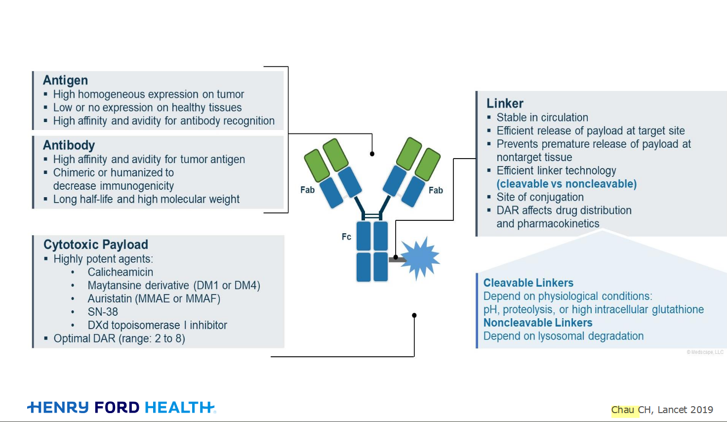
An antibody–drug conjugate (ADC) works a bit like a Trojan horse. It has three main components:

Welcome to the new CancerGRACE.org! Explore our fresh look and improved features—take a quick tour to see what’s new.