
Welcome!
Welcome to the new CancerGRACE.org! Explore our fresh look and improved features—take a quick tour to see what’s new.

Dr. Edward S. Kim from the Levine Cancer Institute in Charlotte, NC defines the concept of cancer histology and gives examples of several lung cancer subtypes.
Transcript
Now we’re talking about histology. When you’ve identified a nodule on a chest x-ray, or a CAT scan, or maybe there’s something in the liver — as you know, lung cancer likes to leave its home base and go to other places — we get a biopsy, and that biopsy is going to help us two ways. One: it’s going to tell us what the origin of the tissue is, and two is: what subtype of that tumor is it? So, in the case of lung cancer, we try to first, identify whether it’s a non-small cell, or small cell lung cancer, and then within the non-small cell lung cancer grouping, which consists of about 85% of all of lung cancer, there are multiple different histology subtypes. That means a pathologist looks at it under a microscope — is looking at it like you would look at artwork on the wall, and trying to identify whether it’s an impressionist period, or it’s a different period of time — and that’s how they’re doing it.
Sometimes, they’ll run some basic tests that they can do in their pathology lab to help further classify one or the other histology subtypes. The most common subtype is adenocarcinoma — again, this is just the name of a non-small cell lung cancer subtype. There are also subtypes called squamous cell cancer, and then — again, those are the two major types, there are then a whole host of others. You will hear terminology such as: large cell carcinoma, neuroendocrine carcinoma, there’s even a classification called NOS, meaning not otherwise specified, and about 10-15% of the time, we can see this.
What does that mean? Well, it still means it’s a lung cancer, and it usually means it’s non-small cell lung cancer, but there is not enough tissue, or the architecture was not preserved enough during the biopsy procedure, that the pathologist can completely classify this tumor. That’s problematic, because now we have therapies that are specifically tailored for some patients who have adenocarcinoma, or squamous cell carcinoma. There are not as many therapies out there tailored for the large cell or neuroendocrine tumors. Again, these just represent different cell types that exist in the lung, and those are the ones that decide to grow and become misbehaving, and they evolve into a cancer, and that’s why they have their particular names.
Please feel free to offer comments and raise questions in our
discussion forums.

Dr. Singhi's reprise on appropriate treatment, "Right patient, right time, right team".
While Dr. Ryckman described radiation oncology as "the perfect blend of nerd skills and empathy".
I hope any...
My understanding of ADCs is very basic. I plan to study Dr. Rous’ discussion to broaden that understanding.
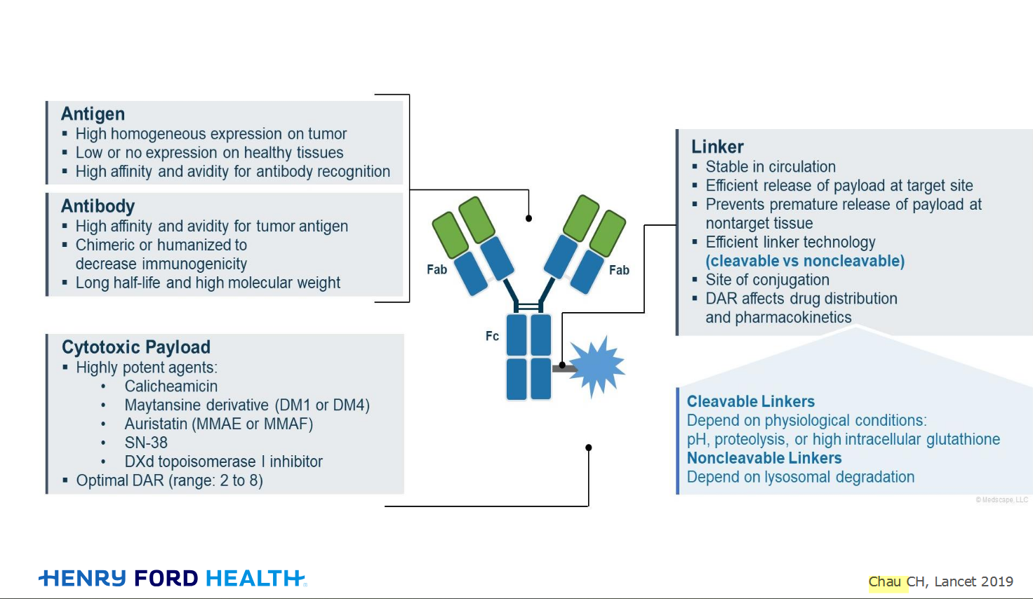
An antibody–drug conjugate (ADC) works a bit like a Trojan horse. It has three main components:
Bispecifics, or bispecific antibodies, are advanced immunotherapy drugs engineered to have two binding sites, allowing them to latch onto two different targets simultaneously, like a cancer cell and a T-cell, effectively...
The prefix “oligo–” means few. Oligometastatic (at diagnosis) Oligoprogression (during treatment)
There will be a discussion, “Studies in Oligometastatic NSCLC: Current Data and Definitions,” which will focus on what we...
Radiation therapy is primarily a localized treatment, meaning it precisely targets a specific tumor or area of the body, unlike systemic treatments (like chemotherapy) that affect the whole body.
The...

Welcome to the new CancerGRACE.org! Explore our fresh look and improved features—take a quick tour to see what’s new.
A Brief Tornado. I love the analogy Dr. Antonoff gave us to describe her presentation. I felt it earlier too and am looking forward to going back for deeper dive.