

Welcome!
Welcome to the new CancerGRACE.org! Explore our fresh look and improved features—take a quick tour to see what’s new.
Transcript
With the pleural fluid that’s built up around the lung — this fluid that exist in between the linings, or as we discussed before, the double plastic bag model — this fluid builds up in there and is trapped, it’s inaccessible. What we need to do is to figure out what that pleural fluid is, and we figure out what that pleural fluid is by sampling it.
What we’re going to talk about today is called thoracentesis, and it’s a really complex term, but it’s really simple in terms of — it is a simple mechanism to get fluid out of the chest and allow us to sort of determine what is going on. So how is this done?
Well, number one: it’s important to figure out that it’s actually pleural fluid that’s causing the symptoms. So, how do we identify people with pleural fluid? We do it based on two basic modalities: one, when symptoms are present, we look for it either with chest x-rays or CT scans, so some of that standard imaging that you may be familiar with, or ultrasound, something that you may not be as familiar with, but this is real-time imaging that allows us, by sticking a simple probe on the back, to see through the chest and into that cavity around the lung and identify if fluid is present.
So once we identify that fluid is present, we need to safely remove that fluid so that we can sample it. So how do we safely do that? The most important thing is that you develop a good relationship with your physician or the person who is going to be draining it, because you need to have confidence — this person is going to be working behind you and talking to you, and you need to sort of develop a comfortable relationship. So take a couple minutes just to talk about what the experience is going to be like, and just sort of get comparable with the whole scenario. Once you’re comfortable, the practitioner may ask you to get ready for a procedure — and how do you get ready for that procedure?
Well, typically what we do is we position people. The most common scenario where people are positioned is sitting on the edge of a bed with your feet on the floor, and leaning over in a position where you may have put a pillow on a table, and laying in front. This has you looking forward, and your physician or practitioner standing behind you, accessing your back. People may also do it with you laying on your back with your arm on your head, giving you access to the side — but these are all different positioning structures so that we can see where the fluid is and most safely access it.
But the point is, is that what we’re going to do together, is get the fluid out safely and comfortably so that you feel better. Your cough should go away, and your shortness of breath should go away, and we should be making a plan for what’s going on for you in the future.
Once you’re positioned, we use the ultrasound machine. Ultrasound has become the standard of care for evaluating whether pleural fluid is present; it allows us to determine what safe access is, where the diaphragm is, where the lung is, where structures that we should avoid are — the best place to go and insert the needle. The next thing that we’re going do it numb up your back. So, to do that, we clean the back, we make sure there’s no risk of infection, then we take a very small needle and insert what’s called, typically, lidocaine. Lidocaine is a medicine that you may have heard of when you go to the dentist and you actually instill a little bit of medicine under the skin — this medicine will be there and it acts very quickly to create a numbing sensation. Then, further and further we go with the needle, deeper, closer to that pleural fluid. Your practitioner should stay in communication with you — “does this hurt, are you feeling okay, does this hurt,” and giving you a little bit more numbing medicine as they go along.
Now, there comes a point where you may experience pain again, that’s the lining of the lung. The lining of that lung, or the outer plastic bag that we talked about, is full of nerve fibers that can be very uncomfortable. So, as the needle touches it, if the numbing medicine hasn’t reached it yet, that can be uncomfortable, so you may want to anticipate that. But then, once you’ve been thoroughly numbed up, that whole track doesn’t feel anything at all, we then slip a very small catheter, far more thin that a pencil, into that pleural sack, and with that we draw the fluid out.
Now, drawing the fluid out can make you cough, and you need to anticipate that because, as the fluid is coming out, the lungs is coming down — fluid come out, lung comes down, but if you think of the lung as a popcorn kernel, as the fluid comes out and the lung is opening up, it can give you this sense to cough. Cough is totally okay, you don't need to worry about that — but if you start experiencing unknown pain, pain in your chest, a deep aching sensation, or tightness, you really need to let your practitioner know.
The other pain that people can feel as the pleural fluid is coming out is pain in your shoulder. So the nerve fibers of the diaphragm that are down here, typically replay back to the shoulder, so if the catheter is touching that or they’re drawing the fluid, you may feel some shoulder pain. That’s okay, just let your practitioner know and they my be able to readjust the catheter.
So, as they’re draining the fluid, they’ll get a lot of feedback information for you — “how are you feeling, are you feeling any pain in there, are you feeling more comfortable,” and the fluid should be able to come out in a very short period of time and you should get clinical relief almost right away. Now, once the fluid has been drained, the catheter is removed, and a simple band aid is placed over it. You should feel free to do almost anything that you want after that. You’re free to go out and eat, you're free to be with your family, you’re free to take a shower. The key things to think about are that, if you have extended pain afterwards or extended shortness of breath, you do need to let your physician or practitioner know about that.
But otherwise, the value of this procedure is to do two things: drain the fluid, with that fluid we get laboratory values — is this fluid infected, does this fluid have cancer in it, do you feel better when I drain it? Not everyone will feel better. But at that point, we need to already start thinking about what we’re going to do in the future for you, because we really don’t want to create an environment where you’re constantly coming back to the office to have to manage this. The goal of this, and the goal for you and your practitioner together, is to maximize your independence, and to move you and liberate you from your frequent office visits as it relates specifically to the fluid that’s building up around the lung.
Please feel free to offer comments and raise questions in our
discussion forums.


A Brief Tornado. I love the analogy Dr. Antonoff gave us to describe her presentation. I felt it earlier too and am looking forward to going back for deeper dive.
Dr. Singhi's reprise on appropriate treatment, "Right patient, right time, right team".
While Dr. Ryckman described radiation oncology as "the perfect blend of nerd skills and empathy".
I hope any...
My understanding of ADCs is very basic. I plan to study Dr. Rous’ discussion to broaden that understanding.
Here's the webinar on YouTube. It begins with the agenda. Note the link is a playlist, which will be populated with shorts from the webinar on specific topics
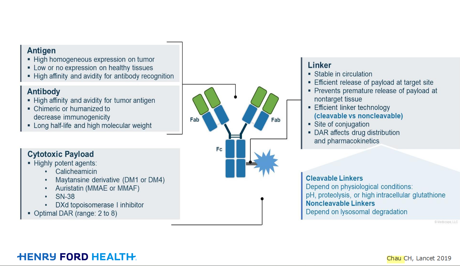
An antibody–drug conjugate (ADC) works a bit like a Trojan horse. It has three main components:
Bispecifics, or bispecific antibodies, are advanced immunotherapy drugs engineered to have two binding sites, allowing them to latch onto two different targets simultaneously, like a cancer cell and a T-cell, effectively...

Welcome to the new CancerGRACE.org! Explore our fresh look and improved features—take a quick tour to see what’s new.
Hi app.92, Welcome to Grace. I'm sorry this is late getting to you. And more sorry your mum is going through this. It's possible this isn't a pancoast tumor even though...