Welcome!
Welcome to the new CancerGRACE.org! Explore our fresh look and improved features—take a quick tour to see what’s new.
We've discussed the potential importance of micrometastatic disease or circulating tumor cells, but another way to assess them is to check for the presence of occult (and microscopic) metastases (OMs) in bone marrow or lymph nodes. The American College of Surgeons Oncology Group recently reported their results on a trial called Z0040 that looked for occult metastases in washings of the pleural space at the time of surgery, from bone marrow collected from a rib, and from lymph nodes that appeared to be negative for cancer involvement by basic pathology review. The key questions were:
1) how common is it to see OMs in the pleural space, marrow, and "negative" lymph nodes?
2) Do patients who test positive for occult metastases (OMs) fare worse than patients who don't have them?
To test this question, from 1999 to 2004, ACOSOG enrolled 1047 patients with resectable stage I-III NSCLC, including 50% with adenocarcinoma, 66% stage I, nearly 50/50 split by patient sex, and median age 67. They underwent surgery in which they underwent "pleural lavage", in which sterile fluid was added to the cavity outside of the lung, then recollected, with a pathologist doing a close search for cancer cells, using immunihistochemistry (IHC), a special staining technique that can identify cells that have proteins consistent with cancer. Patients also had a 3-4 cm piece of rib sent off for bone marrow to be extracted, with a pathologist also doing a careful search for cancer cells, using IHC, and all lymph nodes that were reviewed and found to be negative for cancer involvement by standard evaluation were also reviewed via IHC. All of these tests were done at a single lab at the University of Southern California (USC). Patients were then followed for a minimum of five years to follow their cancer status and survival.
What they found was that OMs in the pleural space or bone marrow were very uncommon (3% and 8%, respectively). The low frequency of OMs in the pleural space made it difficult to say anything meaningful about these patients. For those with OMs in the bone marrow, they showed the same frequency of involvement from stage Ia to IIIB (there were very few patients with stage IIIB NSCLC who underwent surgery, as you would expect) and didn't show evidence of doing worse than those who didn't have OMs in their bone marrow.
The story was different for the patients with OMs in their lymph nodes that were considered negative. Nodal OMs were detected in 22.4% of patients who underwent surgery and were more common in higher stage patients, as you might expect, though they were still seen in about 17% of patients with a smaller, otherwise node-negative cancer. What was so important was that the investigators found that nodal OMs were associated with an approproximately 60% higher risk of developing recurrence and also dying at a given time point over the next 5 years after surgery, compared with those who didn't have nodal OMs, as shown below (top is disease-free survival, and bottom is overall survival):
What does this mean? The authors note that these results are directly relevant to clinical practice, and I agree. We struggle in the post-operative setting with who really needs adjuvant chemotherapy, which is given to eradicate micrometastatic disease. While it is standard to recommend it to people with clearly node-positive disease, the question of which patients with node-negative disease to suggest chemotherapy for remains an open question. Because it can be toxic, giving chemotherapy to people who have a low enough risk of recurrence that they aren't likely to benefit may be worse than just observing them. But it is very easy to imagine that the people with microscopic OMs in their lymph nodes and no other evidence of nodal involvement, who as we see are at considerable risk of recurrence and death from lung cancer, are the patients who might be most likely to benefit from adjuvant chemotherapy. We don't know that with certainty, but the authors note that these IHC and close examination techniques are available through pathology labs everywhere, even if they require a certain level of time and skill to do (I don't know whether the amount of time required is really feasible everywhere). My interpretation is that these results are important enough for me to want to present them for review and discussion with my own multidisciplinary thoracic oncology team, with a thought that we should pursue changing own practices to address these findings and identify patients with nodal disease that might only be negative on first pass. My hope is that this could lead to more refined set of recommendations of which patients are more likely to benefit from adjuvant chemotherapy.
Please feel free to offer comments and raise questions in our
discussion forums.
Dr. Singhi's reprise on appropriate treatment, "Right patient, right time, right team".
While Dr. Ryckman described radiation oncology as "the perfect blend of nerd skills and empathy".
I hope any...
My understanding of ADCs is very basic. I plan to study Dr. Rous’ discussion to broaden that understanding.

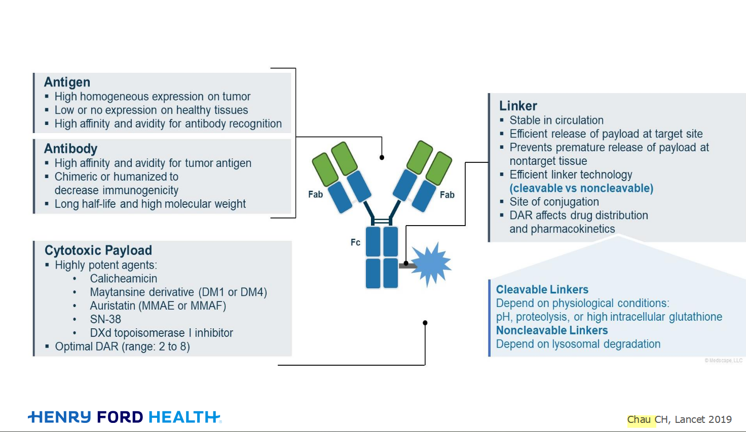
An antibody–drug conjugate (ADC) works a bit like a Trojan horse. It has three main components:
Bispecifics, or bispecific antibodies, are advanced immunotherapy drugs engineered to have two binding sites, allowing them to latch onto two different targets simultaneously, like a cancer cell and a T-cell, effectively...
The prefix “oligo–” means few. Oligometastatic (at diagnosis) Oligoprogression (during treatment)
There will be a discussion, “Studies in Oligometastatic NSCLC: Current Data and Definitions,” which will focus on what we...
Radiation therapy is primarily a localized treatment, meaning it precisely targets a specific tumor or area of the body, unlike systemic treatments (like chemotherapy) that affect the whole body.
The...

Welcome to the new CancerGRACE.org! Explore our fresh look and improved features—take a quick tour to see what’s new.
A Brief Tornado. I love the analogy Dr. Antonoff gave us to describe her presentation. I felt it earlier too and am looking forward to going back for deeper dive.