Welcome!
Welcome to the new CancerGRACE.org! Explore our fresh look and improved features—take a quick tour to see what’s new.
We know PET scans can provide additional metabolic information that can be more sensitive and specific for cancer than chest x-rays and even CT scans in the initial staging of lung cancer (see prior post on introduction to PET scans). PET scans are now nearly universally employed in the initial workup, at least of patients who have NSCLC and aren’t already known to have stage IV disease. But how useful is this technology in the setting of surveillance for the patient at risk for recurrent/residual disease after curative treatment?
Unlike CT scans, which are great at discerning shape and size of internal parts of the body, PET scans are metabolic studies that hold the promise of distinguishing between residual viable cancer and non-viable scar tissue after surgery or radiation. A handful of studies in the post-treatment setting show that PET scans have a 96% sensitivity (PET scans identify a problem 96% of the times that there is a problem) and an 84% specificity (there is viable cancer 84% of the time that the PET scan indicates that it’s there) (example references here and here and here). The problem, though, is that unlike the situation of initial staging, where patients haven’t undergone interventions that could cloud the interpretation of a PET scan, except perhaps for a recent biopsy), after surgery or radiation, the inflammatory changes after treatment can also light up on PET scan and make it difficult to interpret what’s cancer and what’s post-treatment inflammation or infection, especially in the vicinity of the original treated cancer. Because of this concern, which is greatest shortly after local treatment, people have generally recommended that PET scans not be pursued until at least 3-6 months after treatment because of the high risk for false positive results (scan shows abnormalities that aren’t really cancer), and also that any areas that appear suspicious on a PET should be confirmed as cancer with CT imaging and a biopsy before pursuing any interventions against presumed cancer. The concept that PET scans will improve our ability to detect curable recurrences or new cancers hasn’t yet been supported by any evidence of better survival or quality of life in patients.
In the face of such a confusing situation, there’s a lot of variability in what oncologists actually do. As a rule, I don’t obtain regular PET scans on patients after curative surgery or chemoradiation, and I’m particularly reluctant to do so in the first few months after completion of treatment. For my patients with a better prognosis, such as stage I and II NSCLC, I routinely obtain CT scans and will order a PET scan if the CT shows something suspicious (typically I’ll get a PET/CT fusion scan 4-6 weeks after the concerning CT to see if the suspicious area has changed and whether it appears as metabolically active on PET). For stage III patients, in whom the risk of recurrence is higher, I do tend to obtain a PET/CT about once per year, otherwise following with CT scans. My general philosophy is that PET scans can be so sensitive but non-specific, especially after chemoradiation with or without surgery for stage III NSCLC, that changes in the absence of CT findings can too often be an anxiety-laden wild goose chase. Chest CT scans provide a lot of detail, so if there isn’t anything suspicious that has emerged from one CT to the next, I’m quite reassured that anything you might find on a PET scan would have a high chance of being a false positive.
This is an area, though, where we’re gaining more experience, and I really hope and expect that we’ll actually get some study results to help guide or practice patterns. In the meantime, it’s the open frontier (the Wild West if you prefer) in terms of using PET scans for surveillance, with people just following their own rules.
Please feel free to offer comments and raise questions in our
discussion forums.
Hi app.92, Welcome to Grace. I'm sorry this is late getting to you. And more sorry your mum is going through this. It's possible this isn't a pancoast tumor even though...
A Brief Tornado. I love the analogy Dr. Antonoff gave us to describe her presentation. I felt it earlier too and am looking forward to going back for deeper dive.
Dr. Singhi's reprise on appropriate treatment, "Right patient, right time, right team".
While Dr. Ryckman described radiation oncology as "the perfect blend of nerd skills and empathy".
I hope any...
My understanding of ADCs is very basic. I plan to study Dr. Rous’ discussion to broaden that understanding.

Here's the webinar on YouTube. It begins with the agenda. Note the link is a playlist, which will be populated with shorts from the webinar on specific topics
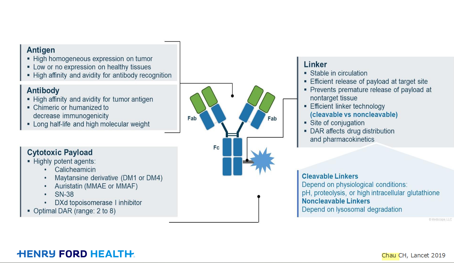
An antibody–drug conjugate (ADC) works a bit like a Trojan horse. It has three main components:

Welcome to the new CancerGRACE.org! Explore our fresh look and improved features—take a quick tour to see what’s new.