Welcome!
Welcome to the new CancerGRACE.org! Explore our fresh look and improved features—take a quick tour to see what’s new.
Purists have considered mediastinoscopy, which is invasive staging of the mediastinum through a small incision just at the base of the neck to get down behind the sternum, or breastbone, to be the "gold standard" for determining whether lymph nodes in the mediastinum, or middle of the chest, is involved with a cancer. The procedure is as shown:
(Click on image to enlarge)
As noted in an earlier post, these lymph nodes are very important in initial staging and also repeat staging after induction therapy; specifically, results after surgery appear to be far superior for patients who have no evidence of residual tumor in the mediastinal nodes after induction therapy, whether chemo or chemo and radiation together. Some thoracic surgeons have a patient undergo mediastinoscopy for initial staging followed by a repeat mediastinoscopy after induction therapy in order to assess response. In fact, that is probably the most definitive way to clarify staging before and after induction treatment. However, mediastinoscopies are not only an invasive procedure but are more complicated when done a second time, and they are also potentially more complicated after treatment, especially if radiation is included. One other potential option for reassessing the mediastinum after surgery is to use imaging, with particular attention to the value of PET scans for determining whether there was a response of the mediastinal lymph nodes.
Just as PET scans provide additional function imaging information to CT scanning for initial staging and potentially also for assessment of response in advanced disease (see my prior post on PET response in advanced NSCLC), some evidence suggests that PET scans can be helpful in providing a non-invasive way to determine whether the mediastinal nodes have responded to induction therapy, which is a potentially important "Go vs. No Go" decision branch point for subsequent surgery or not. This has been evaluated by a few groups. Dr. Cerfolio, a thoracic surgeon from the University of Alabama, has done some interesting early work on this subject. He reported results of a retrospective review of 56 patients with various stages of resectable NSCLC at his institution who had pre-operative chemotherapy or chemo and radiation, who had received a chest CT and PET scan before and after induction therapy (abstract here). He found that the degree of decline in maximum SUV (of disease anywhere, primary mass or mediastinal nodes) on treatment was very inversely correlated with survival (so major decline is very well associated with very good survival, while minimal change was associated with quite poor survival), and this correlation was much better than the change in the size of tumors by CT. Moreover, if the maximum SUV dropped by 90% or more on the repeat PET, there was very close to perfect prediction of a complete pathologic response (no evidence of viable tumor, even under the microscope, in what was taken out in surgery after induction therapy).
Another interesting trial out of Belgium (abstract here) tested the value of CT scans vs. PET scans before and after induction cisplatin-based chemotherapy in 47 patients with stage IIIA N2 NSCLC (of an original 69, the remainder ineligible due to PET detecting higher stage disease than expected in some, and several with "technical/logistical problems"). Some of these patients went on to surgery after the chemo, and the others without progression received radiation. CT findings after induction therapy showed a trend but no significant correlation with survival, but focal uptake in the mediastinum on PET was associated with a worse survival. The separation of node stages by CT and PET, as correlated with survival, are shown here:

This study also looked at PET scan results measured several ways and found that decreases in PET uptake were very highly correlated with long-term survival.
Finally, the group from Belgium, which has led some much of the charge in increasing use of PET scans in lung cancer, did a prospective trial (i.e., planned ahead of time, not just reviewing results after the fact) of PET-CT fusion scans compared directly to repeat mediastinoscopy to see which approach did a better job in predicting outcomes after induction therapy (abstract here). Thirty patients with proven stage IIIA N2 NSCLC received both initial mediastinoscopy and scans, then received 3-4 cycles of cisplatin-based chemo, and then repeat scans and a repeat mediastinoscopy. The PET/CT scans showed no evidence of residual lymph node involvement in 13 patients, N1 (hilar lymph nodes, within the lung) disease in 3, and evidence of persistent N2 node involvement in 14 of the 30 patients. The repeat mediastinoscopy confirmed residual N2 disease in only 5 cases, and 12 cases could not have a good re-mediastinoscopy performed because of scarring in the area. The final surgery on these patients demonstrated that there were actually involved lymph nodes in a total of 17 of these patients. The PET/CT had found 13 of these, and one was a false positive from inflammation. The other four were missed by the PET/CT, three with microscopic areas of residual disease. In contrast, the repeat mediastinoscopy missed 12 patients who had a "false negative" mediastinoscopy but showed mediastinal involvement in the subsequent more extensive surgery. Thus, the overall accuracy of PET/CT was greater than that for re-mediastinoscopy. This trial didn't report survival, so we can't comment on how well either of these approaches predicted outcomes other than the surgical results.
What can we say from these studies about how to proceed in our regular practice for trying to assess how patients have responded to induction chemotherapy or chemoradiotherapy? Nothing definitive. These are small but provocative studies of patients not treated with identical approaches, and in some cases looking at patients of different stages; some received chemo, some chemoradiation. But there is certainly a growing momentum that repeat PET scan results may provide helpful if not perfectly predictive information to be used potentially instead of or in addition to invasive restaging. For some doctors and patients, the emerging evidence will be enough to decide not to pursue a repeat mediastinoscopy. The decision to pursue surgery after induction is a very important one, and we want to ensure that the patients who do it have a feasible possibility of long-term survival. It's not clear whether repeat PET scanning is as accurate if radiation is included in the induction setting, as there is a concern that radiation can induce inflammation that could be picked up as a false positive on PET. We have almost no experience to address these questions right now, but these are the kind of questions we are asking in the field. Additional studies are being conducted, but currently there is at least some growing evidence that PET scan results can potentially add useful information in this setting.
Please feel free to offer comments and raise questions in our
discussion forums.
Dr. Singhi's reprise on appropriate treatment, "Right patient, right time, right team".
While Dr. Ryckman described radiation oncology as "the perfect blend of nerd skills and empathy".
I hope any...
My understanding of ADCs is very basic. I plan to study Dr. Rous’ discussion to broaden that understanding.

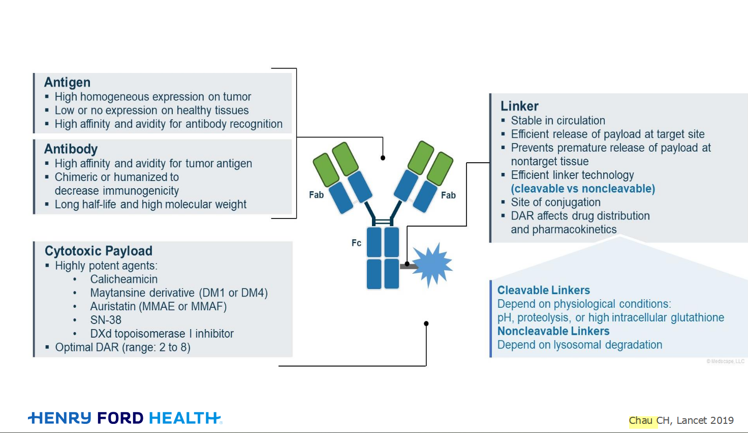
An antibody–drug conjugate (ADC) works a bit like a Trojan horse. It has three main components:
Bispecifics, or bispecific antibodies, are advanced immunotherapy drugs engineered to have two binding sites, allowing them to latch onto two different targets simultaneously, like a cancer cell and a T-cell, effectively...
The prefix “oligo–” means few. Oligometastatic (at diagnosis) Oligoprogression (during treatment)
There will be a discussion, “Studies in Oligometastatic NSCLC: Current Data and Definitions,” which will focus on what we...
Radiation therapy is primarily a localized treatment, meaning it precisely targets a specific tumor or area of the body, unlike systemic treatments (like chemotherapy) that affect the whole body.
The...

Welcome to the new CancerGRACE.org! Explore our fresh look and improved features—take a quick tour to see what’s new.
A Brief Tornado. I love the analogy Dr. Antonoff gave us to describe her presentation. I felt it earlier too and am looking forward to going back for deeper dive.