Welcome!
Welcome to the new CancerGRACE.org! Explore our fresh look and improved features—take a quick tour to see what’s new.
Superior vena cava (SVC) syndrome is an infrequent but not rare complication of lung cancer, occurring in 2-4% of cases, most typically an early symptom that leads to the diagnosis. The SVC is the main vein that drains blood back into the heart from the upper body, and it runs in the middle of the chest on the right side, where it is vulnerable to being compressed by a nearby lung cancer or enlarged lymph nodes, such as from lung cancer or lymphoma. Less commonly, SVC syndrome can be caused by a clot within the blood vessel, and it's also possible to have a combination of external compression and blood clot (clots are more likely to develop where blood flow is compromised). This leads to blockage of the blood flow from the upper body and engorged blood vessels and often swelling of the face, neck, and sometimes upper extremities, as shown in this figure (from this summary article):  The leading symptoms of SVC syndrome are facial edema, distended veins in the neck and sometimes chest, arm edema, shortness of breath, cough, facial plethora/fullness, and less commonly wheezing, lightheadedness, headaches, and even confusion.
Although several decades ago infectious issues such as syphylis and TB were common causes of SVC syndrome, it's much more common now to have cancer as a cause today. Specifically, this is usually lung cancer, but lymphoma can also lead to this, and more rarely causes like germ cell tumors in the chest. A few decades ago, cancer was the source of up to 90% of SVC syndromes, but now that indwelling catheters and pacemakers are more common and can lead to clotting within the blood vessels, cancer is the cause in only about 2/3 of cases. The best way to assess this is with a chest CT with intravenous contrast, although ultrasound studies of the upper extremities (OK, arms) can help identify the extent of the backup; MRI scans are also sometimes used, but generally in people who can't receive IV contrast. The diagnosis is made by obtaining tissue, which is sometimes done by draining pleural fluid, since up to 2/3 of patients with SVC syndrome also have a pleural effusion. Whie a thoracentesis (removing fluid around the lung with a needle from the back) is relatively can provide some relief of shortness of breath, it only yields a diagnosis about half of the time. Bronchoscopy works about 50-70% of the time, CT-guided biopsy with a needle from outside of the chest about 75% of the time, and mediastinoscopy somewhere in the range of 90% of the time. Another potential biopsy source would be to excise an enlarged lymph node, such as above the clavicle, which has the advantage of providing a significant amount of tissue to examine. Particularly for lymphoma but also for lung cancer and other malignant causes, haivng more tissue to review is always helpful. Next, we'll turn to management of SVC syndrome.
Please feel free to offer comments and raise questions in our
discussion forums.
A Brief Tornado. I love the analogy Dr. Antonoff gave us to describe her presentation. I felt it earlier too and am looking forward to going back for deeper dive.
Dr. Singhi's reprise on appropriate treatment, "Right patient, right time, right team".
While Dr. Ryckman described radiation oncology as "the perfect blend of nerd skills and empathy".
I hope any...
My understanding of ADCs is very basic. I plan to study Dr. Rous’ discussion to broaden that understanding.

Here's the webinar on YouTube. It begins with the agenda. Note the link is a playlist, which will be populated with shorts from the webinar on specific topics
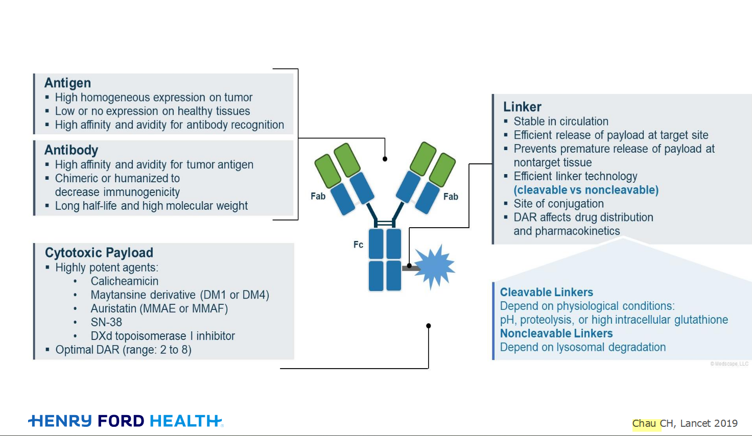
An antibody–drug conjugate (ADC) works a bit like a Trojan horse. It has three main components:
Bispecifics, or bispecific antibodies, are advanced immunotherapy drugs engineered to have two binding sites, allowing them to latch onto two different targets simultaneously, like a cancer cell and a T-cell, effectively...

Welcome to the new CancerGRACE.org! Explore our fresh look and improved features—take a quick tour to see what’s new.
Hi app.92, Welcome to Grace. I'm sorry this is late getting to you. And more sorry your mum is going through this. It's possible this isn't a pancoast tumor even though...