2 Responses to How Long A Period of Follow-Up is Long Enough to Be Confident a Ground Glass Opacity Won’t Grow?
Article and Video CATEGORIES
An interesting article from Japan was published out in the Journal of Thoracic Oncology that asks how long a duration of follow-up imaging of a ground-glass opacity (GGO) is really needed to be confident it's going to remain stable and not grow. It's very common to see small lung nodules that are ambiguous in their significance, for which follow-up scans are typically recommended rather than diving into a biopsy, and non-solid, hazy GGOs are another form of lung lesion that might possibly represent a lung cancer but are also the way a little inflammation or small infection would appear. Even when they turn out to be something technically called cancer based on its appearance under the microscope, it's often a non-invasive adenocarcinoma (sometimes termed bronchioloalveolar carcinoma, or BAC, but shifting in terminology to adenocarcinoma in situ, or AIS) or minimally invasive adenocarcinoma (MIA), in which the invasive component is less than 5 mm in diameter. Even when they grow, it can be at an extremely slow pace.
The report in question looked retrospectively at what happened to 108 GGOs or partially solid nodules (less than 50% solid, more than half hazy and non-solid-appearing on chest CT) in 61 patients, some of whom had a prior history of a resected lung cancer, and some of whom had multiple foci of lung lesions. This wasn't a typical population that we'd define as high risk for lung cancer in North America: 2/3 were women, and 2/3 were never-smokers. Though they included lesions up to 3 cm, nearly 2/3 (64%) were 1 cm or smaller in diameter, and another 31% were 1.1 to 2 cm.
With a median follow-up of 4.2 years, they found that 39 of the 108 lesions either became larger (29) and/or increased in their solid component (17); the others remained stable (within 1 mm) over time, which in some cases went out a decade or longer. Because this was a retrospective review, they didn't have a fixed protocol for when or how to do surgery or other interventions, but over the period of follow-up, 21 patients with 26 lesions underwent surgery. The authors report that in most cases, they turned out to be a pre-malignant lesion like atypical adenomatous hyperplasia (AAH) or AIS or perhaps MIA. Though they don't speak to the survival, the implication is that these patients all did very well, though I suspect the patients who didn't undergo surgery also did extremely well. Notably, 19 of the 21 tumors (90%) tested for an EGFR mutation were positive, further underscoring that this is a very specific population in Japan that can't be readily extrapolated to a global population.
The main point of their report was that all of the lesions that demonstrated any change in size or increased solid component did so within 3 years of the start of follow-up, so that should probably be an appropriate outer limit for regular repeat scans before relaxing some and feeling that it's unlikely the lesion(s) in question will change with longer follow-up. That's consistent with the general guidelines: though there's some variability among the different professional groups, in most cases, 2-3 years of stability is considered to be sufficient to consider a lesion as benign.
What I think is also critically important is the distinction between an extremely slowly changing nodule, even one that is technically called an indolent cancer, and one that is actually a threat to a person's survival over the next few years, or perhaps EVER. What this report shows is that any lesion that doesn't change over three years is clinically insignificant. However, it doesn't say that a lesion that grows by 2 mm over 2.5 years is actually a threat: even if it's called a cancer technically, it might actually only become a problem if it grows over 40 more years.
We don't know that for sure, and our general mindset in cancer management is to treat everything in this realm aggressively. The danger, though, is that as we do more and more chest CTs for lung cancer screening or just because someone coughs once in the emergency room, we're going to find a lot of lung lesions that cause a great deal of anxiety, many follow-up scans, and potentially surgeries for a problem that is only a problem based its relationship as a cousin of a truly threatening cancer. It is important for doctors and patients to recognize the potential for "over-diagnosis" of clinically insignificant things, so that we don't have the treatment be far worse than the underlying disease.
This isn't to say that lung cancer isn't a terrifying disease that takes a heartbreaking toll, but rather that it's important to recognize the biologic variability in how different things that technically all fall under the broad term of "lung cancer" actually behave. We don't want to under-treat or over-treat patients. One important thing about this work is that it highlights that lung nodules can be amazingly stable for years and years, even in some people who have many of them. We may do more harm by going in with guns blazing to treat them.
Please feel free to offer comments and raise questions in our
discussion forums.
Online Community
Hi app.92, Welcome to Grace. I'm sorry this is late getting to you. And more sorry your mum is going through this. It's possible this isn't a pancoast tumor even though...
A Brief Tornado. I love the analogy Dr. Antonoff gave us to describe her presentation. I felt it earlier too and am looking forward to going back for deeper dive.
Dr. Singhi's reprise on appropriate treatment, "Right patient, right time, right team".
While Dr. Ryckman described radiation oncology as "the perfect blend of nerd skills and empathy".
I hope any...
My understanding of ADCs is very basic. I plan to study Dr. Rous’ discussion to broaden that understanding.

Here's the webinar on YouTube. It begins with the agenda. Note the link is a playlist, which will be populated with shorts from the webinar on specific topics
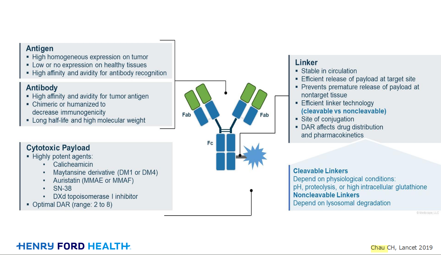
An antibody–drug conjugate (ADC) works a bit like a Trojan horse. It has three main components:
- The antibody, which serves as the “horse,” specifically targets a protein found on cancer...
Bispecifics, or bispecific antibodies, are advanced immunotherapy drugs engineered to have two binding sites, allowing them to latch onto two different targets simultaneously, like a cancer cell and a T-cell, effectively...


hermann says:
Is BAC the only nsclc you should not treat with guns blazing? After my mother thorocotomy may 9, 2013. my 63 yrs old mother was dx with stage IV adeno of the lungs… The cancer is confirmed in three lobes… All the nodules in her lungs were present on a ct scan in 2008. The ct scans in 2013 showed 1cm change in one in the lul and 1 mm change in the nodule in rll. After both were confirm adeno with a needle biopsy, her course of treatment was going to be two thorocotemy to remove both of the confirmed spots of cancer. During surgery they discover the extent of the disease and that it was present in her plural fluid. the docs took a look around the lower left lobe and wedge resect two small adeno. Her lymph nodes are negative and the cancer has not spread outside her lungs? Could opening her up have awaking the sleeping giant? Is it going to start growing out of control? The surgeon told me it was frank adeno… He first thought adeno with BAC features or multifocal stage one adeno. I also want to mention she was asymptomatic before the surgery. I also read a study on beta blockers slowing the growth of lung cancer… could this be slowing it down because she has been on them since 2008. Now 3 weeks after the surgery the docs are talking about putting in a port? Haven’t even seen the oncologist yet.?!? Ask for the pathology report but was told it would only say adeno… Was hoping for markers for target therapy, is that not a standard test for cancer biopsy? Seeing a oncologist soon, can’t wait because I have a ton of questions…
Dr West says:
There’s nothing to say that BAC has a monopoly on indolent growth, and I would put far more emphasis on the pattern of the disease that you’re actually seeing, rather than the diagnosis under the microscope.
The idea that surgery accelerates the cancer is just a prevalent myth, but there’s no evidence at all that this happens. I am not remotely suspicious of that.
Unfortunately, the fact that it was found in the pleural fluid, as well as that it is in a few spots in the lungs, means that it’s beyond a point where cure could be expected, but that doesn’t mean that there’s any clear rationale to treating not only before it is symptomatic but also potentially for a very long time before it becomes symptomatic. Please see this post for a good discussion of this issue
http://cancergrace.org/lung/2013/01/20/mf-bac-algorithm/
This post also clearly states that there is no reason to limit this approach to BAC only. It should apply to all sorts of indolent cancers.
I would say that there’s no very good evidence that beta blockers slow the progression of lung cancer in a meaningful way. Though there are stray studies showing that all sorts of things might slow cancer in lab studies, there is no real evidence that beta blockers have a meaningful impact in real patients.
Good luck.
-Dr. West